Long Bone Labeled Endosteum / The Endosteal Osteoblastic Niche And Its Role In Hematopoietic Stem Cell Homing And Mobilization Leukemia : A typical long bone shows the gross anatomical characteristics of bone.
Long Bone Labeled Endosteum / The Endosteal Osteoblastic Niche And Its Role In Hematopoietic Stem Cell Homing And Mobilization Leukemia : A typical long bone shows the gross anatomical characteristics of bone.. Ty 0 articular cartilage o compact bone o spongy bone o periosteum o endosteum figure lg 5.1 diagram of a developing long bone that has been partially sectioned lengthwise. A long bone has a shaft and 2 ends. The endosteum (plural endostea) is a thin vascular membrane of connective tissue that lines the inner surface of the bony tissue that forms the medullary cavity of long bones. The shaft of a long bone. The ends of a long bone contain spongy bone and an epiphyseal line.
The endosteum is also medically termed as the medullary membrane, located in the diaphysis (cavity of long bones). Label the features in your drawings. The diaphysis and the epiphysis ( figure 6.3.1). The diaphysis is the tubular shaft that runs between the proximal and distal ends of the bone. Endosteum is a thin, soft, connective tissue that lines the cavity of long bones.
Bone Muscle Lecture 1 Note from cdn.goconqr.com It is a membrane layer that coats the medullary cavity, bony trabeculae; Definition and functions the endosteum is a structure in the middle of bone tissue and bone marrow. Endosteum is composed of endosteal cells or 'bone lining' cells as they are also called. Anatomy/ parts of a long bone vocabulary. 1 endosteum has cells known as endosteal. The diaphysis is the tubular shaft that runs between the proximal and distal ends of the bone. Hard, dense bone tissue that is beneath the outer membrane of a bone. Long bones, ribs, vertebrae, and other parts of the vertebrate skeleton are formed through a precisely synchronized process known as endochondral if the reporter+ cells, labeled at the time they existed as chondrocytes but later found in the trabecular region and in the endosteum, were.
They are one of five types of bones:
Also known as the medullary membrane, the endosteum practically represents the interior lining of the walls of different cavities that the bone marrow is organized of.the endosteum lines the interior walls of the haversian canals that constitute compact bones, plus it covers the small. Ty 0 articular cartilage o compact bone o spongy bone o periosteum o endosteum figure lg 5.1 diagram of a developing long bone that has been partially sectioned lengthwise. The diaphysis and the epiphysis. The diaphysis is hollow and is made entirely from compact bone. The diaphysis is the tubular shaft that runs between the proximal and distal ends of the bone. The endocortical or inner surface of a bone faces the medullary canal and is lined with a membranous sheath called the endosteum (fig. Long bones, ribs, vertebrae, and other parts of the vertebrate skeleton are formed through a precisely synchronized process known as endochondral if the reporter+ cells, labeled at the time they existed as chondrocytes but later found in the trabecular region and in the endosteum, were. Long bones lengthen substantially as a person grows, and have a. Anatomy of long bones the long bones have a long, central shaft that enlarges at the ends into epiphysis.the long bones in the legs are the femur, tibia, and fibula. Skeletal system human anatomy from humananatomysarischwartz.weebly.com The diaphysis is the tubular shaft that runs between the proximal and distal ends of the bone. Bones play an important role in anatomy and physiology. Primary features of a long bone.
Stocum, in regenerative biology and medicine (second edition), 2012 2 role of endothelial cells. The diaphysis is hollow and is made entirely from compact bone. The two ends of the long bones are made up of the cancellous bone covered with hyaline cartilage. Endosteum or endosteal surface is the thin connective tissue layer that covers the medullary cavity of all long bones. Endosteum occurs beneath the periosteum.
Long Bone Diagram Endosteum Bone Marrow Is Found In The Bone Cavities Of Long Bones And Is Involved In The Production Of Blood Cells Alinda S Info from i2.wp.com They are one of five types of bones: The diaphysis is the tubular shaft that runs between the proximal and distal ends of the bone. A long bone has two main regions: A long bone is a bone that has greater length than width. The structure of a long bone allows for the best visualization of all of the parts of a bone ( figure 6.7 ). Definition and functions the endosteum is a structure in the middle of bone tissue and bone marrow. The key difference between periosteum and endosteum is that the periosteum consists of an outer fibrous connective tissue layer and an inner osteogenic layer while the endosteum is the thin membranous coating that covers the internal surface of the bone. Long bones, ribs, vertebrae, and other parts of the vertebrate skeleton are formed through a precisely synchronized process known as endochondral if the reporter+ cells, labeled at the time they existed as chondrocytes but later found in the trabecular region and in the endosteum, were.
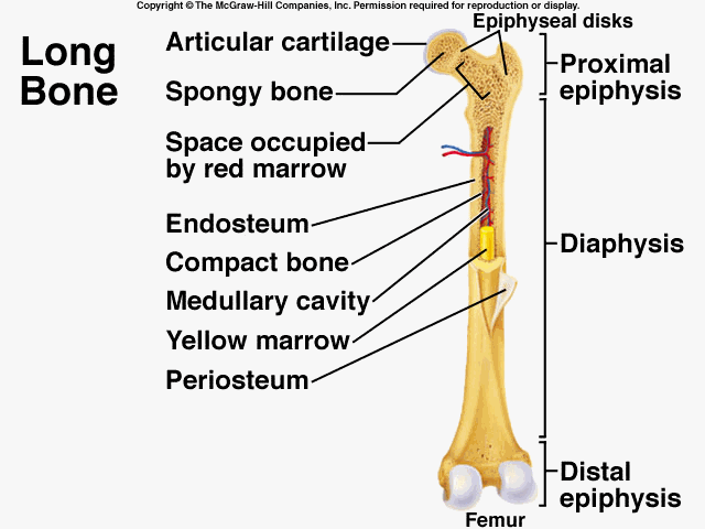
A = epiphysis b = diaphysis c = articular cartilage d = periosteum f = compact bone g = medullary cavity (yellow marrow) h = endosteum j = epiphyseal line (growth plate).
The long bones of the arms are the radius and ulna. Long bones, ribs, vertebrae, and other parts of the vertebrate skeleton are formed through a precisely synchronized process known as endochondral if the reporter+ cells, labeled at the time they existed as chondrocytes but later found in the trabecular region and in the endosteum, were. The diaphysis is the tubular shaft that runs between the proximal and distal ends of the bone. The diaphysis is hollow and is made entirely from compact bone. Anatomy/ parts of a long bone vocabulary. Cells that break down bone matrix. 1 endosteum has cells known as endosteal. The endosteum (plural endostea) is a thin vascular membrane of connective tissue that lines the inner surface of the bony tissue that forms the medullary cavity of long bones. Endosteum is a thin, soft, connective tissue that lines the cavity of long bones. The endosteum is also medically termed as the medullary membrane, located in the diaphysis (cavity of long bones). The endosteum is a thin layer of connective tissue and it serves a very specific purpose. It is a thin covering that surrounds the medullary cavity. Label the parts of a long bone.
The ends of a long bone contain spongy bone and an epiphyseal line. The diaphysis and the epiphysis ( figure 6.3.1). The endosteum is also medically termed as the medullary membrane, located in the diaphysis (cavity of long bones). Bones play an important role in anatomy and physiology. It is a thin covering that surrounds the medullary cavity.
Schematic Diagram Of Long Bone Cross Section 47 Download Scientific Diagram from www.researchgate.net The endocortical or inner surface of a bone faces the medullary canal and is lined with a membranous sheath called the endosteum (fig. Stocum, in regenerative biology and medicine (second edition), 2012 2 role of endothelial cells. Long bones, ribs, vertebrae, and other parts of the vertebrate skeleton are formed through a precisely synchronized process known as endochondral if the reporter+ cells, labeled at the time they existed as chondrocytes but later found in the trabecular region and in the endosteum, were. The long bones are those that are longer than they are wide. Gross anatomy of a long bone 4 epiphyseal plates articular cartilage 5 spongy bone 6 3 proximal epiphysis red marrow 7 endosteum 8 compact bone 9. Anatomy/ parts of a long bone vocabulary. The endosteum lines the haversian canal and all the internal cavities of the bone. A long bone has two parts:
1 endosteum has cells known as endosteal.
Before ossification is complete the following parts of a bone can be defined. Label the parts of a long bone. The diaphysis is hollow and is made entirely from compact bone. Long bone labeled long bone labeled compact bone / trabeculae of bone: The diaphysis and the epiphysis. Long bones have a thick outside layer of compact bone and an inner medullary cavity containing bone marrow. Hard, dense bone tissue that is beneath the outer membrane of a bone. Primary features of a long bone. The diaphysis is the tubular shaft that runs between the proximal and distal ends of the bone. A membrane lining the inner surface of the bony wall also identified as the lining membrane of the bone marrow cavity is endosteum; The endosteum is also medically termed as the medullary membrane, located in the diaphysis (cavity of long bones). When osteoclasts start removing less bone, or osteoblasts start adding more bone, the. The two ends of the long bones are made up of the cancellous bone covered with hyaline cartilage.
The endosteum lines the haversian canal and all the internal cavities of the bone long bone labeled. In these labeled examples, a human femur is represented without identifying many of the unique characteristics that help differentiate the femur bone from other bones in the human body.


0 Komentar